16+
DOI: 10.18413/2658-6533-2025-11-1-0-1

Pathway directed mechanisms of anti-EGFR resistance in colorectal cancer (review)

Abstract

Background: Targeting the epidermal growth factor receptor (EGFR) with cetuximab or panitumumab (anti-EGFR MAb) has been historically reserved for patients with RAS/BRAF wild-type advanced colorectal cancer (CRC). However, results of recent studies including PARADIGM and PRESSING evaluating the role of negative hyperselection of RAS wild-type CRC by alterations in other genes suggest that other genomic factors beyond RAS/BRAF/ERBB2 might influence the response to anti-EGFR MAbs in CRC. Although vast, current data on the predictive role of individual biomarkers to anti-EGFR MAb is often misunderstood. The aim of the study:In this review, in light of recent findings, we aimed to summarize existing data on the influence of various signaling pathways and their individual components along with nongenomic factors for the optimal patient selection for anti-EGFR MAbs. Materials and methods: To collect available information on possible mechanisms of resistance to anti-EGFR MAb in patients with colorectal cancer we searched PubMed and ClinicalTrials.gov in May 2024. We also searched proceedings from the major oncology conferences ESMO, ASCO, and ASCO GI up to May 2024. We further scanned reference lists from eligible publications. Results: In this review we outline current knowledge on the mechanisms of resistance to anti-EGFR MAbs beyond traditional KRAS/NRAS/BRAF mutations in CRC. We focus on the alterations of genes involved in signaling pathways downstream of EGFR that can be detected by comprehensive tumor profiling in real-world clinical practice. Conclusion: Despite many mechanisms affecting various signaling pathways beyond the traditional KRAS/NRAS/BRAF mutations that are thought to be implicated in the resistance to anti-EGFR MAb in CRC, future efforts are needed to clarify their significance. Ongoing sequencing efforts will clarify the need for expanding the list of alterations routinely tested for the selection of candidates for anti-EGFR MAb


Introduction. Advanced colorectal cancer (CRC) is the third most commonly diagnosed cancer, and one of the leading causes of cancer-related deaths [1]. Many molecular mechanisms of CRC progression have been described, including genes involved in Ras/Raf/MEK/ERK known as the mitogen-activated protein kinases (MAPK) pathway, the phosphatidylinositol 3-kinase (PI3K)/protein kinase B (AKT) pathway as well as Wnt/β-catenin, transforming growth factor-β1/SMAD (TGF-β/SMAD) and Janus kinase/signal transducer and activator of transcription 3 (JAKs/STAT3) pathways [2]. Concomitantly genes regulating metabolism, as well as genes responsible for biotransformation of xenobiotics and antioxidant enzymes etc., affect the effectiveness of anti-EGFR MAb therapy, reducing it [3].

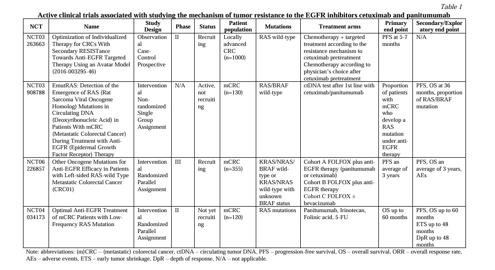
Development and approval of anti-EGFR monoclonal antibodies (MAb), cetuximab and panitumumab, have provided a significant survival benefit for patients with KRAS/NRAS (RAS) and BRAF wild type (wt) CRC [4]. However, while the development of acquired resistance to treatment is inevitable in most patients, some patients demonstrate intrinsic resistance [5-8]. In current clinical practice, treatment decisions regarding anti-EGFR MAbs are based on the analysis of classic biomarkers of resistance, such as mutations in RAS and BRAF genes [9, 10]. Various nonsystemic studies have previously reported on the influence of individual genomic alterations beyond RAS/RAF mutations on the benefit of anti-EGFR MAb therapy. However, the results of PRESSING and PARADIGM trials have truly reopened the question of the optimal selection of CRC patients for the anti-EGFR MAbs. Unlike previous studies, these studies suggest that the simultaneous screening of various genes frequently upregulated in CRC might be the most effective approach [11, 12]. However, current data suggests that not all of the alterations might be the same in terms of influencing the resistance to therapy. Therefore, it is important to address the impact of individual alterations in various genes, as the genes included in diagnostic panels used in real-world clinical practice might vary. In addition, several clinical trials are still ongoing (Table 1). In this review, we discuss current understanding of the mechanisms of resistance to anti-EGFR MAbs in CRC beyond RAS/BRAF V600 mutations. Here, we review mechanisms of both primary and acquired resistance with a focus on altered signaling pathways, and not on differentiation between the two.

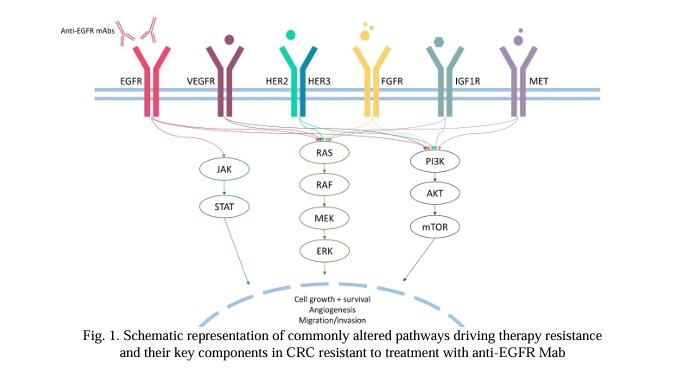
1. Unconventional mechanisms of RAS/RAF-mediated resistance. The mitogen-activated protein kinases (MAPK) pathway, often known as the Ras/Raf/MEK/ERK signaling pathway, is a highly conserved signal transduction pathway in all cells. The MAPK pathway is one of the best-characterized signaling cascades that regulates cell proliferation, differentiation, survival and apoptosis, by transmitting signals from upstream extracellular growth factors to various downstream effectors [13] (Fig.1). However, although widely investigated, there are still some unanswered questions regarding RAS/RAF-mediated resistance.

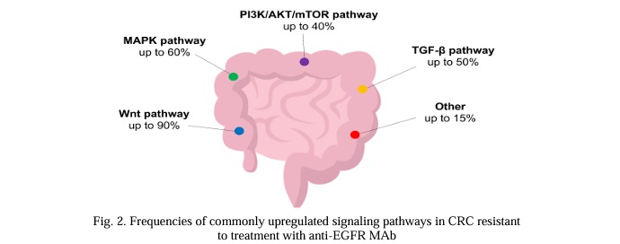
1.1 Unanswered questions regarding RAS. Oncogenic mutations in RAS are the most common molecular event in CRC (Fig.2). RAS mutations are detected in up to 60% of CRC patients; KRAS mutations are found in around 30% of cases, while NRAS mutations are found in about 5% of cases [14, 15]. Activating mutations are predominantly located in 2, 3 and 4 exons, affecting the catalytic G domain. The majority of observed mutations are substitutions that occur in hotspots affecting codons 12 (70-80% of all cases), 13 and 61 [16]. Rare activating missense variants affect codons 59, 117 and 146 [17]. Oncogenic activation of RAS genes is a known mechanism of resistance to anti-EGFR MAbs in CRC patients [18], as shown in numerous clinical studies [19-24]. RAS mutations are routinely analyzed by PCR, and the implementation of NGS analysis may increase the detection rate by 9% with tissue and X ctDNA analysis, respectively [25].

Although KRAS mutations are validated predictive biomarkers of resistance to cetuximab and panitumumab, patients harboring the KRAS G13D mutation might still derive benefit from cetuximab, as shown in several retrospective studies [26, 27]. This effect may be attributed to the atypical activating effect of the variant, as shown in functional studies [28]. A possible mechanism that distinguishes KRAS G13D from other activating variants was shown using a mathematical model and biochemical studies [29, 30]. According to the model, KRAS G13D is most likely sensitive to cetuximab due to a difference in the mechanism of interaction with NF1 and wt RAS. While other KRAS variants bind to wt RAS negative regulator NF1, effectively inhibiting the wt RAS inhibitor and leading to wt HRAS and NRAS activation, G13D does not interact with NF1, thus promoting NF1-mediated inhibition of wt RAS and the effective functioning of cetuximab. Thus, cetuximab treatment can block wt HRAS and NRAS activation. Although this hypothetical mechanism explains the atypical effect of the KRAS G13D variant, its clinical relevance remains unknown. Despite this evidence, a more recent retrospective analysis indicates that patients with KRAS G13D mutations are unlikely to respond to cetuximab [31]. However, both large retrospective and prospective trials failed to confirm this effect. Teipar et al. [27] reported a significant improvement of PFS (median, 7.4 vs 6.0 months; hazard ratio [HR], 0.47; P = 0.039) and tumor response (40.5% v 22.0%; odds ratio, 3.38; P = 0.042), but not survival (median, 15.4 vs 14.7 months; HR, 0.89; P = 0.68) in those receiving cetuximab harboring KRAS codon 13 mutations. Similarly, in a retrospective analysis by Peeters et al. [32] the presence of KRAS G13D was significantly associated with a negative impact on OS (P = 0.0018). In a phase II Fleming single-stage design study by Schirripa et al. [33] evaluating the activity of single-agent cetuximab in KRAS G13D-mutated CRC, the primary objective of the trial was not met, DCR at 6 months was 0%.

Studies show that patients with low variant allele frequencies (VAF) of RAS/RAF might still be candidates for anti-EGFR therapy. In a post hoc analysis of the CRYSTAL study, it was shown that patients with RAS-mutated CRC whose mutations had low VAF in the tumor (0.1%-5%) benefited from the addition of cetuximab to FOLFIRI [34]. Similar results were obtained in a phase II ULTRA trial. Based on the results of this study, the optimal threshold for VAF of RAS/RAF mutations was established to be 5%. Across patients with RAS/BRAF mutation whose mutations had VAF of 5% and less, the response rate as well as median overall survival (OS) and progression-free survival (PFS) were similar to the RAS/BRAF wt patient cohort [35]. Noteworthy, this threshold is only used when tissue is analyzed and is not applicable for ctDNA analysis. When ctDNA is used, the tumor is considered RAS-mutant if the mutation is identified with any VAF.

Another member of the RAS oncogene family is HRAS. However, alterations in HRAS are rare in CRC [15], hence only anecdotal evidence points toward the association between HRAS mutations and resistance to anti-EGFR MAbs [36].

In the case of KRAS G12C-mutated CRC, another treatment strategy is the combination of specific KRAS G12C inhibitors (i.e. sotorasib, adagrasib) to anti-EGFR MAbs [37]. The combination treatment is preferable to KRAS G12C inhibitor monotherapy, as frequently observed mechanisms of acquired resistance to these drugs include new RTK pathway alterations, which can be targeted by EGFR inhibitors [38].

Finally, KRAS amplification is another alteration that can be found in CRC. KRAS amplifications are extremely rare among patients with CRC (<1%) and are usually mutually exclusive with other KRAS alterations [39]. KRAS amplification has been suggested to drive resistance to anti-EGFR treatment in a small patient cohort [18, 39, 40]. In vitro cetuximab could partially abrogate phosphorylation of MEK and ERK but, like in KRAS mutant cells, was unable to induce growth arrest in KRAS amplification-positive cells [18].

1.2 De novo RAS mutations and neoRAS. Another known mechanism of acquired resistance to anti-EGFR MAbs is the emergence of de novo RAS mutations in the course of treatment [18, 41, 42, 43]. The emergence of RAS mutant subclones can be detected months prior to the radiographic documentation of disease progression via liquid biopsy [18, 43]. At the same time, the discontinuation of anti-EGFR MAb might be associated with a decrease of the level of acquired RAS mutations [44]. Thus, assessment of acquired RAS mutations in liquid biopsies is necessary not only for the timely detection of acquired resistance to EGFR inhibitors, but also for when considering therapy re-challenge [45, 46, 47].

An opposite phenomenon known as ‘neoRAS wild-type’ is characterized by the conversion of RAS-mutant tumors to RAS wt, as detected in ctDNA in the course of treatment with standard therapies. However, this phenomenon is thought to be uncommon, occurring in only 1-8% of patients [48]. One patient in the case series by Osumi et al. [49] with neoRAS has been reported to exhibit a long-term partial response (PR) to panitumumab in combination with irinotecan. Results of the SCRUM-Japan GOZILA study reported an incidence of neoRAS of 9%. In this study, out of 6 patients with neoRAS, 1 patient had PR and another had SD for at least 6 months [50].

1.3 Non-V600 BRAF mutations. Activating mutations in BRAF occur in 8-12% of patients with CRC, with the most common missense mutation BRAF V600E accounting for up to 95% of all BRAF mutations [51-54]. BRAF V600E is widely known to be one of the most common causes of primary resistance to EGFR therapy in CRC [55, 56]. BRAF V600E leads to constitutive activation of the MAPK pathway, and therefore inhibition of the MAPK pathway by EGFR inhibitors alone is not effective [57]. However, it has also been shown that monotherapy with BRAF inhibitors is also not effective in BRAFV600E CRC, which may be explained by EGFR-mediated feedback reactivation of MAPK signaling [58]. The addition of BRAF inhibitors in combination with EGFR inhibitors has been shown to restore the sensitivity of BRAF-mutant tumors [59-65]. A combination of encorafenib and cetuximab was approved by the FDA for the treatment of patients with BRAF V600E CRC based on the results of the BEACON trial [62].

Class II and III BRAF variants can be seen in about 2.2% of CRC patients. BRAF non-V600 mutations mostly affect codons 594 and 596 [66, 67, 68]. Class II mutations are activating and signal in dimers in a RAS-independent manner. Class III BRAF mutations typically exhibit reduced kinase activity or absence thereof, however can still activate MAPK through signaling through increased RAS binding or CRAF activation, which is RAS-dependent [69].

The data on the effect of non-V600 BRAF mutations in terms of their influence on the efficacy of anti-EGFR therapy is conflicting [70]. Preclinical data as well as several case reports suggest that BRAF non-V600 mutations (specifically, class IIB and III mutations) may be sensitive to EGFR inhibition due to the dependency on upstream receptor tyrosine kinase signaling [51, 71].

However, clinical studies have suggested that BRAF non-V600 mutations might be implicated in the resistance to anti-EGFR MAb. In a retrospective study among 36 patients with BRAF class II and III mutations, the median survival of patients was significantly higher than for patients with BRAF V600E (36.1 months vs 21 months), however, among 11 patients receiving anti-EGFR therapy, no responses were observed, while 6 patients achieved stable disease as best response [72]. Similar results were obtained in another study, where among 4 CRC patients with non-V600 BRAF variants no one responded to cetuximab therapy [73]. Studies suggest that different non-V600 BRAF mutations might have a different effect on the efficacy of anti-EGFR MAb. In a study by Yaeger et al. [74] it has been suggested that response in CRCs with class II BRAF mutants is rare, while a large portion of CRCs with class III BRAF mutants might respond to therapy, based on the difference in objective response rate (ORR) in the two groups (8% vs 50%).

Differences in the effect of anti-EGFR MAbs on CRC with BRAF mutations of different classes may be attributed to differences in their biological properties. For instance, class III BRAF mutations are largely dependent on upstream EGFR signaling, and thus might be more sensitive to EGFR inhibition [71]. Additionally, non-V600 BRAF mutations in CRC are rare, and thus may be largely understudied.

1.4 Other mutations in MAPK pathway genes. Mutations in genes other than RAS/BRAF in the MAPK pathway may also be associated with resistance to anti-EGFR MAbs in CRC.

For example, gain of function mutations of MAP2K1 (encoding for MEK1) have been suggested as one of potential drivers of primary resistance but are not recommended for routine assessment due to insufficient validation in clinical trials [75]. MAP2K1 mutations, especially p.Lys57, were found in CRC patients with shorter PFS [76, 77] and were also recently found to be implicated in acquired resistance to anti-EGFR agents [39, 75, 78].

NF1, another gene involved in the MAPK pathway, encodes for a negative regulator of KRAS and plays a negative regulatory role in signaling downstream of EGFR due to its function as a RAS GTPase activating protein [79, 80]. It was demonstrated that NF1 loss might be one of the potential mechanisms of acquired resistance to EGFR inhibitors in CRC [76, 81, 82]. NF1 inactivation has also been associated with decreased sensitivity of human lung cancer cells to EGFR inhibitors, which can be attributed to enhanced RAS signaling [83].

GTPase RAC1 and its alternatively spliced isoform RAC1B, important components of the pathobiology of various tumor progression processes, were shown to be involved in anti-EGFR MAb resistance using CRC cell lines [84], as well as surgical specimen from head and neck squamous cell carcinoma (HNSCC) patients [85].

Mutations in ARID1A, the most frequently mutated subunit of the SWI/SNF chromatin remodeling complex in cancer, have been reported to be associated with a transcriptional signature predicting reduced efficacy of anti-EGFR MAbs. This effect can be partially attributed to the activation of PI3K/MAPK signaling and loss of SWI/SNF activity [86]. However, further studies are warranted to confirm these findings.

2. PI3K pathway-mediated resistance. The PI3K/AKT/mTOR (PI3K) pathway is the second most commonly upregulated intracellular signaling pathway in CRC. In CRC, the oncogenic activation of the PI3K pathway frequently occurs through gain of function mutations of PIK3CA, as well as loss of function mutations, deletions or loss of expression of PTEN – all common events in CRC [87]. For instance, PIK3CA exon 20/PTEN/AKT1 alterations were found in 10.9% of older patients receiving panitumumab plus FOLFOX or 5-FU/LV [88]. The PI3K pathway is an important signaling pathway downstream of EGFR, exhibiting crosstalk with other signaling pathways, including MAPK [89, 90]. It has been proposed that the oncogenic activation of the PI3K pathway might play a role in generating resistance to EGFR-targeting therapies in CRC due to the activation of signaling downstream of EGFR [89, 91]. However, since dysregulation of the PI3K pathway often coexists with RAS/BRAF mutations, the individual roles of PI3K alterations in terms of anti-EGFR resistance warrants further investigation [87].

2.1 PIK3CA. Oncogenic mutations in PIK3CA occur in RAS-mutant and in RAS-wt CRC, suggesting that they might possess both driver and passenger roles depending on the molecular context [92, 93]. PIK3CA is altered in up to 20% of CRCs [87, 92] (Fig.2). In over 1.5% of tumors, double-hit mutations are observed, which are associated with increased PI3Kα signaling [94]. Activating mutations are predominantly located in exons 9 and 20 of the gene, affecting kinase and helical domains. However, recent studies suggest that less common activating mutations can occur in other exons of the gene [95, 96, 97].

Although molecular testing is routinely performed for patients with CRC, the data on the activity of anti-EGFR MAbs against PIK3CA-mutated tumors is limited. Although responses can be observed occasionally, PIK3CA mutations are generally associated with resistance to anti-EGFR MAbs, as shown by lower PFS and OS rates across patients with PIK3CA mutations [25, 91, 98, 99]. This effect appears to be especially pronounced in patients with exon 20 PIK3CA mutations, whereas exon 9 mutations do not seem to be predictive of response to anti-EGFR therapy [91, 99, 100]. This difference can be attributed to the domain-specific nature of activating properties of various PIK3CA mutations [101]. However, some studies report no effect of PIK3CA mutations on OS in RAS wt tumors [98, 102]. Although potentially significant, these findings should be interpreted with caution, since no randomized controlled trials have been carried out.

2.2 PTEN. When compared to PIK3CA, PTEN is less frequently altered in CRC (~5-7%) [87]. PTEN is a negative regulator of the PI3K pathway, and its loss or loss of function leads to aberrant PI3K signaling [103]. PTEN loss of function (LoF) mutations, as well as loss of protein expression due to promoter hypermethylation are associated with features of the sessile-serrated pathway [104, 105]. PTEN mutations/loss of expression have been associated with reduced response rates to cetuximab [106, 107]. Lack of response to panitumumab has also been reported across patients with PTEN loss or LoF mutations [107]. Additionally, reduced PTEN copy number has also been implicated in resistance [106, 107]. However, the number of studies that investigated the individual effect of PTEN alterations is small, warranting further validation.

2.3 Other components of the PI3K pathway. Although mutations affecting PIK3CA or PTEN are the most common in CRC, impact of alterations in other genes encoding for the components of the PI3K/Akt/mTOR pathway has been reported. PIK3R1 encodes the p85α subunit of PI3K and acts as a regulator of the p110α catalytic product of the PIK3CA locus. Additionally, it has been proposed that PIK3R1, together with PIK3R2, is involved in the regulation of PTEN protein stability [108, 109]. Interestingly, PIK3R1 tends to be altered in RAS/BRAF wt tumors, albeit at low frequency [76]. In vitro studies have identified decrease of PIK3R1 expression as a potential mechanism of anti-EGFR resistance [110].

Point mutations in AKT1 occur at lower rates as compared to PIK3CA/PTEN alterations in CRC [86]. AKT1 oncogenic mutations, primarily E17K, had been identified in CRC patients initially resistant to anti-EGFR treatment [111, 112]. Additionally, AKT1 mutations are associated with concurrent RAS/BRAF mutations [111].

The impact of FBXW7 alterations on the resistance to anti-EGFR MAbs remains controversial. The F-box protein FBXW7 is also implicated in the PI3K signaling [113]. Alterations of FBXW7 have been identified in CRC patients displaying short PFS and lack of response to anti-EGFR treatment, however the small sample size and the retrospective nature of data do not allow to draw univocal conclusions [76, 114, 115].

Combinations of anti-EGFR MAbs with various targeted agents, such as mTOR inhibitors, have also resulted in high efficacy, warranting further validation in larger patient cohorts [116, 117, 118]. However, clinical evaluation of an experimental PI3K inhibitor combined with cetuximab for KRAS wt CRC patients unselected for the alterations of PI3K/Akt/mTOR pathway, had limited activity [119].

3. RTK. Mutations in receptor tyrosine kinases (RTK) lead to the autophosphorylation of the tyrosine kinase domain resulting in conformation changes and activation of downstream signaling pathways. Alterations of different RTKs can be found in CRC, the majority of which have the potential to activate PI3K and MAPK signaling [120], highlighting that the oncogene addiction can be a driver of anti-EGFR therapy resistance.

3.1 HER/ERBB family. Amplifications of ERBB2 occur in up to 3% of tumors, and outline a distinct patient population [87]. RAS/BRAF/PIK3CA quadruple wild-type tumors are especially enriched for ERBB2 amplifications, which are observed in up to 20-30% of cases [121]. ERBB2 amplifications have long been recognized as a mechanism of primary resistance to anti-EGFR mAbs, as they have been shown to be associated with worse PFS and ORR in various studies [122, 123]. However, the question of what threshold of ERBB2 amplification should be taken into account when considering EGFR MAb requires further studies [124].

In a similar manner to ERBB2 amplifications, ERBB2 mutations result in downstream pathway activation, and thus have the potential to mediate resistance to EGFR-targeting agents [125]. Consistently, various studies have validated the association between ERBB2 mutations, particularly the ones occurring in the tyrosine kinase of the protein, and resistance to EGFR inhibitors. Interestingly, ERBB2 mutations can be attributed to both primary and acquired resistance [126]. In the era of NGS, these findings have become more relevant than ever before due to the potential of NGS to identify not only ERBB2 amplifications, but also mutations.

Preclinical studies suggest that ERBB3 mutations may also influence the effectiveness of EGFR MAb in CRC, as well as in HNSCC. This effect can be explained by the activation of the PI3K pathway caused by ERBB3 oncogenic mutations [127]. However, in a retrospective study by Loree et al. ERBB3 mutations exhibited a less pronounced effect on the effects of EGFR MAb treatment when compared to ERBB2 mutations [128].

For cancers with sustained ERBB signaling, the addition of ERBB TKIs has been investigated, resulting in promising antitumor activity [129, 130, 131]. Despite the promising activity of other agents, when combined with cetuximab or panitumumab, pan-ERBB TKI neratinib failed to produce any objective responses among patients with RAS/BRAF/PIK3CA wt CRC [132].

3.2 EGFR ECD mutations. In some cases, acquired resistance to anti-EGFR MAbs can be mediated by the emergence of EGFR ectodomain (ECD) point mutations [133]. EGFR ECD mutations may arise in up to 16% of patients treated with EGFR MAbs [133]. Several EGFR ECD mutations have been reported, including, among others, R451C, S492R, G465R, K467T [134-138]. Contrary to activating mutations of the EGFR, most of the EGFR ECD mutations lie in the surface recognized by EGFR MAbs and have the potential to affect complex formation. Some mutations (i.e. R451C) that are not specifically located in EGFR MAb binding sites introduce other critical structural changes [134]. Importantly, a subset of these mutations (such as S492R) may only affect the interaction with cetuximab and not panitumumab [133, 134], which may be attributed to the presence of a large central cavity in panitumumab but not cetuximab [139].

Switching from cetuximab to panitumumab has been reported to be an effective strategy for treating cetuximab-resistant CRC patients with acquired EGFR ECD mutations, as a significant subset of these mutations does not prevent panitumumab binding [134]. Moreover, Sym004, a 1:1 mixture of cetuximab and panitumumab has been shown to be an effective treatment strategy for EGFR ECD-mutated CRC in vitro [140]. Therefore, for CRC patients with EGFR ECD mutations no additional agents might be needed apart from standard EGFR MAb.

3.3 MET amplification. Alterations of the MET are a well-established mechanism of resistance to EGFR tyrosine kinase inhibitors (TKIs) in non-small cell lung cancer (NSCLC) [141, 142]. Similarly to NSCLC, MET amplifications, albeit occurring at lower frequencies (around 1%), have been found to drive resistance to anti-EGFR mAb in CRC. Using patient-derived tumor xenografts, Badrelli et al. showed that MET amplification might be associated both with primary and acquired resistance in KRAS wt CRC, which was then supported by patient cases [143]. This effect can be attributed by the activation of the downstream PI3K and MAPK induced by MET [144].

In MET-amplified CRC, a combination of MET and EGFR targeting agents have resulted in the improvement of patients’ outcomes, although the data on the combination of these drugs are limited [145].

3.4 Kinase fusions. Kinase fusions are rare in CRC, occurring in less than 1% of the patients [87, 146]. However, kinase gene fusions are estimated to be enriched in RAS/BRAF wt tumors in patients that will be potentially treated with EGFR MAbs [147], or mismatch repair deficient/microsatellite unstable (dMMR/MSI) tumors [148, 149]. Specifically, ALK, BRAF, NTRK, RET gene fusions have been reported in patients with CRC [88, 112, 147, 150]. Kinase gene fusions have been identified in EGFR MAb treatment resistant CRC patients [112], however, their effect has been underexplored in large randomized studies due to low prevalence. Additionally, kinase fusions have been found to be enriched in dMMR/MSI colorectal cancer, which can further explain resistance [151].

3.6 Other. Other uncommon RTK-mediated mechanisms of resistance have been suggested. For instance, although rare in CRC, in vitro studies have identified FGFR1 amplifications as mediating resistance to anti-EGFR mAbs, possibly due to the activation of compensatory pathways, however, these findings have not been further investigated to date [143].

Upon binding with growth factors and insulin, the insulin-like growth factor 1 receptor (IGF1R) activates the two most commonly upregulated signaling pathways in CRC, the PI3K and MAPK [152]. Elevated expression of IGF1R has been found to be a poor prognostic factor in CRC [153]. Low IGF1R expression has been found to correlate with better outcomes with cetuximab treatment [154]. However, the data regarding the effect of IGF1R expression on the efficacy of EGFR-targeting therapy is inconsistent [154, 155].

Persistent activation of the JAK/STAT pathway has also been linked to anti-EGFR therapy. Specifically, activation of STAT3 through phosphorylation correlated with the resistance to EGFR TKI gefitinib in CRC cell lines, suggesting that STAT3 phosphorylation may play a role in mediating resistance to EGFR inhibitors in CRC [156].

VEGF and EGFR signaling pathways are closely related, sharing multiple downstream effectors. VEGF signaling plays a crucial role in angiogenesis, and inhibition of angiogenesis is one of the mechanisms of action of anti-EGFR MAb. Increased expression of VEGF has been reported to be associated with decreased response to cetuximab [157]. Dual targeting of VEGF and EGFR represents a promising strategy for overcoming resistance mediated by either VEGF, or VEGF/EGFR crosstalk [158, 159].

5. Wnt signaling and epithelial-to-mesenchymal transition. The Wnt pathway is commonly divided into β-catenin dependent, or canonical, and independent, or non-canonical, signaling pathways [160]. The canonical Wnt signaling pathway plays one of the most important roles in CRC carcinogenesis [161]. Wnt activation in CRC occurs through inactivation of APC approximately in 50% of cases, or through mutation of β-catenin [160]. Wnt signaling pathway promotes the nuclear accumulation of β-catenin, which contributes to epithelial-mesenchymal transition (EMT) and increased tumor aggressiveness [162]. In CRC cell cultures, it was shown that the expression of E-cadherin, a marker of epithelial cells, may be associated with the effectiveness of EGFR inhibition. At the same time, mesenchymal cells were 7 times less sensitive to anti-EGFR MAbs when compared to epithelial cells [163]. These findings were also validated in other tumor types. For instance, in treatment-naive patients with HNSCC who received cetuximab before surgery, upregulation of expression of genes implicated in CAF and EMT including markers of embryologic pathways like NOTCH and Wnt was demonstrated [164]. There is also supporting preclinical data for other cancer types besides CRC [165-168].

While some studies suggest that APC mutations might contribute to anti-EGFR MAb resistance, the data is inconsistent. For instance, Thota et al. reported that APC mutations in the context of TP53 mutations may, in fact, predict cetuximab sensitivity [169].

6. TP53. Alterations in TP53, commonly referred to as ‘guardian of the genome’ can be found in the vast majority of CRC cases (>70%) [87]. Several studies have reported that TP53 wild-type or TP53-expressing CRC tumors exhibit worse outcomes when treated with anti-EGFR MAbs when compared to TP53-mutant tumors, however other factors, such as tumor sidedness, might contribute to this effect [170-173]. In vitro studies suggest that EGFR expression can be differently modulated depending on the TP53 mutational status, and TP53-mutant status is generally associated with increased EGFR expression, which can explain the differences in anti-EGFR MAb sensitivity [174, 175].

7. TGF-β pathway. The transforming growth factor (TGF)-β signaling pathway is involved in many biologic cellular processes such as cell proliferation, differentiation, apoptosis, and extracellular matrix production [176]. In the early CRC carcinogenesis, activation of TGF-β leads to tumor suppression [177]. However, in advanced stages, TGF-β is believed to promote metastasis, angiogenesis, and EMT [178, 179].

SMAD4 is a common mediator in the transcriptional regulator complex in the TGF-β pathway [180]. It has been demonstrated that SMAD4 mutations can lead to cetuximab resistance in CRC patients. The modified PFS (mPFS) and ORR to cetuximab has been reported to be decreased for SMAD4-mutated patients when compared to SMAD4 wt patients [81, 115]. Similar results have also been shown in other studies [181] and for other tumor types, specifically for HNSCC [182, 183].

8. Non-genomic mechanisms of resistance. Various other non-genomic mechanisms driving EGFR MAb resistance have been proposed. For instance, dMMR, caused by a dysfunction of mismatch repair and occurring in up to 15% of CRC patients, has been reported to play an important role in mediating resistance [184]. Although the mechanism underlying resistance in dMMR/MSI CRC cases remains largely unknown, it has been shown that in cases where dMMR/MSI is caused by the hypermethylation of MLH1 promoter (commonly referred to as sporadic dMMR/MSI) increase in the expression of ERBB2, as well as PI3K signaling can be observed [185]. However, in this case, the increase in the ERBB2 expression might not be clinically significant, as ERBB2 amplification-positivity and MSI are mutually exclusive in CRC [186]. Additionally, sporadic dMMR/MSI is commonly associated with BRAF V600E mutations, whereas mutations in other oncogenes known to drive resistance to anti-EGFR MAb are frequently found in Lynch syndrome-associated CRC [187].

Apart from dMMR/MSI, the immune microenvironment by itself can be considered as an important modulator of anti-EGFR MAb efficacy. Specifically, increase in cancer-associated fibroblasts (CAFs), which produce mitogenic growth factors such as FGF1, FGF2, HGF, TGF-β and others, as well as angiogenesis and abnormal functioning of various immune cells have been demonstrated to modulate resistance [82].

Additionally, metabolic reprogramming, which can occur following treatment with EGFR-targeting agents, can influence therapy efficacy [188]. Autophagy, a self-cannibalization biological process, is another factor that should be considered when discussing mechanisms of resistance to EGFR-targeting agents. It has also been proposed that autophagy acts as a protective response in cancer cells [189]. Finally, cancer stem cells, also known as tumor-initiating cells, have also been suggested to play a part in drug resistance, due to their ability to self-renew and differentiate into various cell lineages [190].

Furthermore, gut microbiota composition has been found to modulate therapy efficacy in various tumors, including CRC. However, data regarding the effect of gut microbiota on the efficacy of anti-EGFR MAbs is currently limited. A small study by Lewandowski et al. [191] found that patients with high diversity of gut microbiome may be better candidates for anti-EGFR MAb therapy, as compared to patients with non-diverse microbiome. However, these findings should be further validated, as diverse gut microbiomes have been linked to a good prognosis of CRC patients regardless of therapy [192].

Finally, non-coding RNAs (ncRNAs), including microRNAs, long non-coding RNAs, and circular RNAs have been reported to regulate resistance to anti-EGFR MAb in CRC. One of the suggested mechanisms explaining this phenomenon is that different ncRNAs can upregulate oncogenic signaling pathways promoting resistance to anti-EGFR therapy [193].

Discussion and conclusions. Monoclonal antibodies that target EGFR blocking downstream signaling have emerged as important therapeutic agents in the treatment of CRC. The indication of drugs from this class, cetuximab and panitumumab, is currently based on the mutational statuses of KRAS, NRAS and BRAF genes [9, 10]. Until recently, the question of whether additional factors play a role in resistance to anti-EGFR MAb has not been widely investigated. However, the encouraging results of trials evaluating the role of the hyperselection have reopened the question of the optimal selection of CRC patients for the anti-EGFR MAbs once again [11, 12].

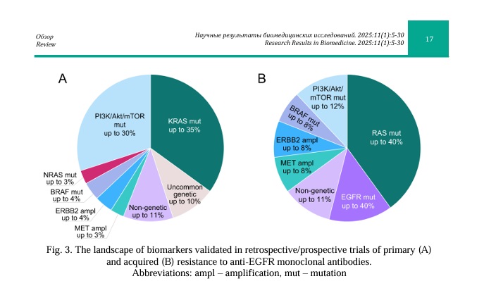
Here, we outline current knowledge on the mechanisms of resistance to anti-EGFR MAbs beyond traditional alterations in CRC. Specifically, we focus on the alterations of various genes involved in relevant signaling pathways downstream of EGFR that can be detected by genomic assays currently used in the real-world clinical practice. Despite the fact that some of the discussed biomarkers have been extensively studied, their individual use in the clinic is limited by the lack of randomized clinical studies. Therefore, it will be necessary to validate the clinical utility of these alterations in large cohort studies for optimal patient management. Future efforts should be directed at optimizing strategies to overcome resistance to anti-EGFR MAbs in patients with various genomic alterations. The most common mechanisms of intrinsic and acquired resistance are summarized in Figure 3.

Many attempts have been made towards the development of strategies to overcome resistance to anti-EGFR MAb in CRC [194]. Combining anti-EGFR MAbs with targeted agents represents a promising strategy for overcoming treatment resistance arising from compensatory pathway activation, although further studies are warranted to improve patient outcomes. Additionally, novel agents may be used as standalone therapies for patients with various alterations, such as aberrant Wnt/β-catenin signaling, for instance [195, 196].

Apart from genetically-driven resistance, growing evidence suggests that various non-genetic mechanisms might be implicated in the resistance to anti-EGFR MAbs in CRC. Although currently these findings are mostly of academic interest, with the advances of novel assays, it will be possible to incorporate their analysis into routine clinical practice.

In current clinical practice, tumor sidedness plays a crucial role in clinical decision making, which is largely driven by differences in their biology. For instance, right-sided tumors are more likely to have MSI, as well as display higher rates of oncogenic EGFR activation, BRAF and PIK3CA mutations, which factor into therapy resistance [197]. However, further efforts should be directed towards the implementation of comprehensive genomic testing into routine clinical practice, which will allow to focus on genomic portraits of tumors, and not only their sidedness.

In conclusion, while many mechanisms affecting various signaling pathways beyond the traditional RAS/BRAF mutations are thought to be implicated in the resistance to anti-EGFR therapy in colorectal cancer, future efforts are needed to clarify their significance. Ongoing sequencing efforts will clarify the need for expanding the list of alterations routinely tested for the selection of candidates for anti-EGFR therapy.

Financial support

This study was supported by a grant from theMoscow Center of Innovative Technologies in Healthcare (№2102-2/23).

Reference lists

  1. Siegel RL, Wagle NS, Cercek A, et al. Colorectal cancer statistics, 2023. CA: A Cancer Journal for Clinicians. 2023;73(3):233-254. DOI: https://doi.org/10.3322/caac.21772
  2. Malki A, ElRuz RA, Gupta I, et al. Molecular Mechanisms of Colon Cancer Progression and Metastasis: Recent Insights and Advancements. International Journal of Molecular Sciences. 2020;24;22(1):130. DOI: https://doi.org/10.3390/ijms22010130
  3. Zhou J, Ji Q, Li Q. Resistance to anti-EGFR therapies in metastatic colorectal cancer: underlying mechanisms and reversal strategies. Journal of Experimental & Clinical Cancer Research. 2021;18;40(1):328. DOI: https://doi.org/10.1186/s13046-021-02130-2
  4. Sun H, Li Y, Su Y, et al. Efficacy and safety of anti-EGFR monoclonal antibodies combined with different chemotherapy regimens in patients with RAS wild-type metastatic colorectal cancer: A meta-analysis. Journal of Evidence-Based Medicine. 2019;12(4):300-312. DOI: https://doi.org/10.1111/jebm.12360
  5. Amado RG, Wolf M, Peeters M, et al. Wild-type KRAS is required for panitumumab efficacy in patients with metastatic colorectal cancer. Journal of Clinical Oncology. 2008;26(10):1626-34. DOI: https://doi.org/10.1200/JCO.2007.14.7116. Corrected and republished in: Journal of Clinical Oncology. 2023;41(18):3278-3286
  6. Van Cutsem E, Köhne CH, Hitre E, et al. Cetuximab and chemotherapy as initial treatment for metastatic colorectal cancer. The New England Journal of Medicine. 2009;360(14):1408-17. DOI: https://doi.org/10.1056/NEJMoa0805019
  7. Douillard JY, Siena S, Cassidy J, et al. Randomized, phase III trial of panitumumab with infusional fluorouracil, leucovorin, and oxaliplatin (FOLFOX4) versus FOLFOX4 alone as first-line treatment in patients with previously untreated metastatic colorectal cancer: the PRIME study. Journal of Clinical Oncology. 2010;28(31):4697-705. DOI: https://doi.org/10.1200/JCO.2009.27.4860
  8. Peeters M, Price TJ, Cervantes A, et al. Randomized phase III study of panitumumab with fluorouracil, leucovorin, and irinotecan (FOLFIRI) compared with FOLFIRI alone as second-line treatment in patients with metastatic colorectal cancer. Journal of Clinical Oncology. 2010;28(31):4706-13. DOI: https://doi.org/10.1200/JCO.2009.27.6055
  9. Benson AB, Venook AP, Adam M, et al. NCCN Clinical Practice Guidelines in Oncology (NCCN Guidelines®) Colon Cancer Version 2.2024 — April 30, 2024 https://www.nccn.org/professionals/physician_gls/pdf/colon.pdf
  10. Cervantes A, Adam R, Roselló S, et al. Metastatic colorectal cancer: ESMO Clinical Practice Guideline for diagnosis, treatment and follow-up. Annals of Oncology. 2023;34(1):10-32. DOI: https://doi.org/10.1016/j.annonc.2022.10.003
  11. Morano F, Corallo S, Lonardi S, et al. Negative Hyperselection of Patients With RAS and BRAF Wild-Type Metastatic Colorectal Cancer Who Received Panitumumab-Based Maintenance Therapy. Journal of Clinical Oncology. 2019;37(33):3099-3110. DOI: https://doi.org/10.1200/JCO.19.01254
  12. Watanabe J, Muro K, Shitara K, et al. Panitumumab vs Bevacizumab Added to Standard First-line Chemotherapy and Overall Survival Among Patients With RAS Wild-type, Left-Sided Metastatic Colorectal Cancer: A Randomized Clinical Trial. JAMA. 2023;329(15):1271-1282. Erratum in: JAMA. 2023;329(24):2196. DOI: https://doi.org/10.1001/jama.2023.4428
  13. Dhillon AS, Hagan S, Rath O, et al. MAP kinase signalling pathways in cancer. Oncogene. 2007;26(22):3279-90. DOI: https://doi.org/10.1038/sj.onc.1210421
  14. Bos JL. ras oncogenes in human cancer: a review. Cancer Research. 1989;49(17):4682-9. Erratum in: Cancer Research 1990;50(4):1352
  15. Fernández-Medarde A, Santos E. Ras in cancer and developmental diseases. Genes Cancer. 2011 Mar;2(3):344-58. DOI: https://doi.org/10.1177/1947601911411084
  16. Pantsar T. The current understanding of KRAS protein structure and dynamics. Computational and Structural Biotechnology Journal. 2019;18:189-198. DOI: https://doi.org/10.1016/j.csbj.2019.12.004
  17. Smith G, Bounds R, Wolf H, et al. Activating K-Ras mutations outwith 'hotspot' codons in sporadic colorectal tumours - implications for personalised cancer medicine. British Journal of Cancer. 2010;102(4):693-703. DOI: https://doi.org/10.1038/sj.bjc.6605534
  18. Misale S, Yaeger R, Hobor S, et al. Emergence of KRAS mutations and acquired resistance to anti-EGFR therapy in colorectal cancer. Nature. 2012;486(7404):532-6. DOI: https://doi.org/10.1038/nature11156
  19. Douillard JY, Oliner KS, Siena S, et al. Panitumumab-FOLFOX4 treatment and RAS mutations in colorectal cancer. The New England Journal of Medicine. 2013;369(11):1023-34. DOI: https://doi.org/10.1056/NEJMoa1305275
  20. Bokemeyer C, Köhne CH, Ciardiello F, et al. FOLFOX4 plus cetuximab treatment and RAS mutations in colorectal cancer. European Journal of Cancer. 2015;51(10):1243-52. DOI: https://doi.org/10.1016/j.ejca.2015.04.007
  21. Peeters M, Oliner KS, Price TJ, et al. Analysis of KRAS/NRAS Mutations in a Phase III Study of Panitumumab with FOLFIRI Compared with FOLFIRI Alone as Second-line Treatment for Metastatic Colorectal Cancer. Clinical Cancer Research. 2015;21(24):5469-79. DOI: https://doi.org/10.1158/1078-0432.CCR-15-0526
  22. Kim TW, Elme A, Kusic Z, et al. A phase 3 trial evaluating panitumumab plus best supportive care vs best supportive care in chemorefractory wild-type KRAS or RAS metastatic colorectal cancer. British Journal of Cancer. 2016;115(10):1206-1214. DOI: https://doi.org/10.1038/bjc.2016.309
  23. Venook AP, Niedzwiecki D, Lenz HJ, et al. Effect of First-Line Chemotherapy Combined With Cetuximab or Bevacizumab on Overall Survival in Patients With KRAS Wild-Type Advanced or Metastatic Colorectal Cancer: A Randomized Clinical Trial. JAMA. 2017;317(23):2392-2401. DOI: https://doi.org/10.1001/jama.2017.7105
  24. Cremolini C, Antoniotti C, Lonardi S, et al. Activity and Safety of Cetuximab Plus Modified FOLFOXIRI Followed by Maintenance With Cetuximab or Bevacizumab for RAS and BRAF Wild-type Metastatic Colorectal Cancer: A Randomized Phase 2 Clinical Trial. JAMA Oncology. 2018;4(4):529-536. DOI: https://doi.org/10.1001/jamaoncol.2017.5314
  25. Vidal J, Bellosillo B, Santos Vivas C, et al. Ultra-selection of metastatic colorectal cancer patients using next-generation sequencing to improve clinical efficacy of anti-EGFR therapy. Annals of Oncology. 2019;30(3):439-446. DOI: https://doi.org/10.1093/annonc/mdz005
  26. De Roock W, Jonker DJ, Di Nicolantonio F, et al. Association of KRAS p.G13D mutation with outcome in patients with chemotherapy-refractory metastatic colorectal cancer treated with cetuximab. JAMA. 2010;304(16):1812-20. DOI: https://doi.org/10.1001/jama.2010.1535
  27. Tejpar S, Celik I, Schlichting M, et al. Association of KRAS G13D tumor mutations with outcome in patients with metastatic colorectal cancer treated with first-line chemotherapy with or without cetuximab. Journal of Clinical Oncology. 2012;30(29):3570-7. DOI: https://doi.org/10.1200/JCO.2012.42.2592
  28. Hunter JC, Manandhar A, Carrasco MA, et al. Biochemical and Structural Analysis of Common Cancer-Associated KRAS Mutations. Molecular Cancer Research. 2015;13(9):1325-35. DOI: https://doi.org/10.1158/1541-7786.MCR-15-0203
  29. McFall T, Diedrich JK, Mengistu M, et al. A systems mechanism for KRAS mutant allele-specific responses to targeted therapy. Science Signaling. 2019;12(600):eaaw8288. DOI: https://doi.org/10.1126/scisignal.aaw8288
  30. McFall T, Stites EC. A mechanism for the response of KRASG13D expressing colorectal cancers to EGFR inhibitors. Molecular & Cellular Oncology. 2020;7(2):1701914. DOI: https://doi.org/10.1080/23723556.2019.1701914
  31. Gajate P, Sastre J, Bando I, et al. Influence of KRAS p.G13D mutation in patients with metastatic colorectal cancer treated with cetuximab. Clinical Colorectal Cancer. 2012;11(4):291-6. DOI: https://doi.org/10.1016/j.clcc.2012.02.003
  32. Peeters M, Douillard JY, Van Cutsem E, et al. Mutant KRAS codon 12 and 13 alleles in patients with metastatic colorectal cancer: assessment as prognostic and predictive biomarkers of response to panitumumab. Journal of Clinical Oncology. 2013;31(6):759-65. DOI: https://doi.org/10.1200/JCO.2012.45.1492
  33. Schirripa M, Loupakis F, Lonardi S, et al. Phase II study of single-agent cetuximab in KRAS G13D mutant metastatic colorectal cancer. Annals of Oncology. 2015;26(12):2503. DOI: https://doi.org/10.1093/annonc/mdv385
  34. Van Cutsem E, Lenz HJ, Köhne CH, et al. Fluorouracil, leucovorin, and irinotecan plus cetuximab treatment and RAS mutations in colorectal cancer. Journal of Clinical Oncology. 2015;33(7):692-700. DOI: https://doi.org/10.1200/JCO.2014.59.4812
  35. Santos C, Azuara D, Viéitez JM, et al. Phase II study of high-sensitivity genotyping of KRAS, NRAS, BRAF and PIK3CA to ultra-select metastatic colorectal cancer patients for panitumumab plus FOLFIRI: the ULTRA trial. Annals of Oncology. 2019;30(5):796-803. DOI: https://doi.org/10.1093/annonc/mdz082
  36. Boidot R, Chevrier S, Julie V, et al. HRAS G13D, a new mutation implicated in the resistance to anti-EGFR therapies in colorectal cancer, a case report. Int J Colorectal Dis. 2016;31(6):1245-6. DOI: https://doi.org/10.1007/s00384-015-2448-7
  37. Fakih MG, Salvatore L, Esaki T, et al. Sotorasib plus Panitumumab in Refractory Colorectal Cancer with Mutated KRAS G12C. The New England Journal of Medicine. 2023;389(23):2125-2139. DOI: https://doi.org/10.1056/NEJMoa2308795
  38. Li BT, Velcheti V, Price TJ, et al. Largest evaluation of acquired resistance to sotorasib in KRAS p.G12C-mutated non–small cell lung cancer (NSCLC) and colorectal cancer (CRC): Plasma biomarker analysis of CodeBreaK100. Journal of Clinical Oncology. 2022;40:102–102 DOI: https://doi.org/10.1200/JCO.2022.40.16_suppl.102
  39. Valtorta E, Misale S, Sartore-Bianchi A, et al. KRAS gene amplification in colorectal cancer and impact on response to EGFR-targeted therapy. International Journal of Cancer. 2013;133(5):1259-65. DOI: https://doi.org/10.1002/ijc.28106
  40. Fang T, Liang T, Wang Y, et al. An Early-Onset Advanced Rectal Cancer Patient With Increased KRAS Gene Copy Number Showed A Primary Resistance to Cetuximab in Combination With Chemotherapy: A Case Report. Frontiers in Oncology. 2021;11:755578. DOI: https://doi.org/10.3389/fonc.2021.755578
  41. Siravegna G, Mussolin B, Buscarino M, et al. Clonal evolution and resistance to EGFR blockade in the blood of colorectal cancer patients. Nature Medicine. 2015;21(7):795-801. DOI: https://doi.org/10.1038/nm.3870. Epub 2015 Jun 1. Erratum in: Nature Medicine. 2015;21(7) DOI: https://10.1038/nm0715-827b. Erratum in: Nature Medicine. 2015;21(7):827.
  42. Parikh AR, Corcoran RB. Monitoring resistance through liquid biopsy. Annals of Oncology. 2018;29(1):8-11. DOI: https://doi.org/10.1093/annonc/mdx650
  43. Siena S, Sartore-Bianchi A, Garcia-Carbonero R, et al. Dynamic molecular analysis and clinical correlates of tumor evolution within a phase II trial of panitumumab-based therapy in metastatic colorectal cancer. Annals of Oncology. 2018;29(1):119-126. DOI: https://doi.org/10.1093/annonc/mdx504
  44. Parseghian CM, Loree JM, Morris VK, et al. Anti-EGFR-resistant clones decay exponentially after progression: implications for anti-EGFR re-challenge. Annals of Oncology. 2019;30(2):243-249. DOI: https://doi.org/10.1093/annonc/mdy509
  45. Cremolini C, Rossini D, Dell'Aquila E, et al. Rechallenge for Patients With RAS and BRAF Wild-Type Metastatic Colorectal Cancer With Acquired Resistance to First-line Cetuximab and Irinotecan: A Phase 2 Single-Arm Clinical Trial. JAMA Oncology. 2019;5(3):343-350. DOI: https://doi.org/10.1001/jamaoncol.2018.5080
  46. Ohhara Y, Shinozaki E, Osawa H, et al. Liquid biopsy for optimizing the rechallenge of cetuximab in metastatic colorectal cancer: Additional study of E-Rechallenge Trial. Journal of Clinical Oncology. 2009;37:585–585 DOI: https://doi.org/10.1200/JCO.2019.37.4_suppl.585
  47. Sunakawa Y, Nakamura M, Ishizaki M, et al. RAS Mutations in Circulating Tumor DNA and Clinical Outcomes of Rechallenge Treatment With Anti-EGFR Antibodies in Patients With Metastatic Colorectal Cancer. JCO Precision Oncology. 2020;4:898-911. DOI: https://doi.org/10.1200/PO.20.00109
  48. Henry J, Willis J, Parseghian CM, et al. NeoRAS: Incidence of RAS reversion from RAS mutated to RAS wild type. Journal of Clinical Oncology. 2020;38:180–180 DOI: https://doi.org/10.1200/JCO.2020.38.4_suppl.180
  49. Osumi H, Vecchione L, Keilholz U, et al. NeoRAS wild-type in metastatic colorectal cancer: Myth or truth?-Case series and review of the literature. European Journal of Cancer. 2021;153:86-95. DOI: https://doi.org/10.1016/j.ejca.2021.05.010
  50. Osumi H, Shinozaki E, Nakamura Y, et al. NeoRAS wild-type metastatic colorectal cancer in the SCRUM-Japan GOZILA study. Journal of Clinical Oncology; 2023;Vol. 41, Issue 16_suppl, pp. 3506–3506). American Society of Clinical Oncology (ASCO). https://doi.org/10.1200/jco.2023.41.16_suppl.3506
  51. De Roock W, Claes B, Bernasconi D, et al. Effects of KRAS, BRAF, NRAS, and PIK3CA mutations on the efficacy of cetuximab plus chemotherapy in chemotherapy-refractory metastatic colorectal cancer: a retrospective consortium analysis. The Lancet Oncology. 2010;11(8):753-62. DOI: https://doi.org/10.1016/S1470-2045(10)70130-3
  52. Davies H, Bignell GR, Cox C, et al. Mutations of the BRAF gene in human cancer. Nature. 2002 Jun 27;417(6892):949-54. DOI: https://doi.org/10.1038/nature00766
  53. Cantwell-Dorris ER, O'Leary JJ, Sheils OM. BRAFV600E: implications for carcinogenesis and molecular therapy. Molecular Cancer Therapeutics. 2011;10(3):385-94. DOI: https://doi.org/10.1158/1535-7163.MCT-10-0799
  54. Barras D, Missiaglia E, Wirapati P, et al. BRAF V600E Mutant Colorectal Cancer Subtypes Based on Gene Expression. Clinical Cancer Research. 2017;23(1):104-115. DOI: https://doi.org/10.1158/1078-0432.CCR-16-0140
  55. Bokemeyer C, Van Cutsem E, Rougier P, et al. Addition of cetuximab to chemotherapy as first-line treatment for KRAS wild-type metastatic colorectal cancer: pooled analysis of the CRYSTAL and OPUS randomised clinical trials. European Journal of Cancer. 2012;48(10):1466-75. DOI: https://doi.org/10.1016/j.ejca.2012.02.057
  56. Pietrantonio F, Petrelli F, Coinu A, et al. Predictive role of BRAF mutations in patients with advanced colorectal cancer receiving cetuximab and panitumumab: a meta-analysis. European Journal of Cancer. 2015;51(5):587-94. DOI: https://doi.org/10.1016/j.ejca.2015.01.054
  57. Xu T, Wang X, Wang Z, et al. Molecular mechanisms underlying the resistance of BRAF V600E-mutant metastatic colorectal cancer to EGFR/BRAF inhibitors. Therapeutic Advances in Medical Oncology. 2022;14:17588359221105022. DOI: https://doi.org/10.1177/17588359221105022
  58. Corcoran RB, Ebi H, Turke AB, et al. EGFR-mediated re-activation of MAPK signaling contributes to insensitivity of BRAF mutant colorectal cancers to RAF inhibition with vemurafenib. Cancer Discovery. 2012;2(3):227-35. DOI: https://doi.org/10.1158/2159-8290.CD-11-0341
  59. Di Nicolantonio F, Martini M, Molinari F, et al. Wild-type BRAF is required for response to panitumumab or cetuximab in metastatic colorectal cancer. Journal of Clinical Oncology. 2008;26(35):5705-12. DOI: https://doi.org/10.1200/JCO.2008.18.0786
  60. Yaeger R, Cercek A, O'Reilly EM, et al. Pilot trial of combined BRAF and EGFR inhibition in BRAF-mutant metastatic colorectal cancer patients. Clinical Cancer Research. 2015;21(6):1313-20. DOI: https://doi.org/10.1158/1078-0432.CCR-14-2779
  61. van Geel RMJM, Tabernero J, Elez E, et al. A Phase Ib Dose-Escalation Study of Encorafenib and Cetuximab with or without Alpelisib in Metastatic BRAF-Mutant Colorectal Cancer. Cancer Discovery. 2017;7(6):610-619. DOI: https://doi.org/10.1158/2159-8290.CD-16-0795
  62. Kopetz S, Grothey A, Yaeger R, et al. Encorafenib, Binimetinib, and Cetuximab in BRAF V600E-Mutated Colorectal Cancer. The New England Journal of Medicine. 2019;381(17):1632-1643. DOI: https://doi.org/10.1056/NEJMoa1908075
  63. Huijberts SC, van Geel RM, Bernards R, et al. Encorafenib, binimetinib and cetuximab combined therapy for patients with BRAFV600E mutant metastatic colorectal cancer. Future Oncology. 2020;16(6):161-173. DOI: https://doi.org/10.2217/fon-2019-0748
  64. Ros J, Baraibar I, Sardo E, et al. BRAF, MEK and EGFR inhibition as treatment strategies in BRAF V600E metastatic colorectal cancer. Therapeutic Advances in Medical Oncology. 2021;13:1758835921992974. DOI: https://doi.org/10.1177/1758835921992974
  65. Tabernero J, Grothey A, Van Cutsem E, et al. Encorafenib Plus Cetuximab as a New Standard of Care for Previously Treated BRAF V600E-Mutant Metastatic Colorectal Cancer: Updated Survival Results and Subgroup Analyses from the BEACON Study. Journal of Clinical Oncology. 2021;39(4):273-284. DOI: https://doi.org/10.1200/JCO.20.02088
  66. Cremolini C, Di Bartolomeo M, Amatu A, et al. BRAF codons 594 and 596 mutations identify a new molecular subtype of metastatic colorectal cancer at favorable prognosis. Annals of Oncology. 2015;26(10):2092-7. DOI: https://doi.org/10.1093/annonc/mdv290
  67. Jones JC, Renfro LA, Al-Shamsi HO, et al. Non-V600 BRAF Mutations Define a Clinically Distinct Molecular Subtype of Metastatic Colorectal Cancer. Journal of Clinical Oncology. 2017;35(23):2624-2630. DOI: https://doi.org/10.1200/JCO.2016.71.4394
  68. Van Cutsem E, Dekervel J. Not All BRAF-Mutant Metastatic Colorectal Cancers Are Identical: Distinct Clinical Consequences of non-V600 BRAF Mutations. Journal of Clinical Oncology. 2017;35(23):2598-2599. DOI: https://doi.org/10.1200/JCO.2017.72.7057
  69. Owsley J, Stein MK, Porter J, et al. Prevalence of class I-III BRAF mutations among 114,662 cancer patients in a large genomic database. Experimental Biology and Medicine (Maywood, N.J.). 2021;246(1):31-39. DOI: https://doi.org/10.1177/1535370220959657
  70. Caputo F, Santini C, Bardasi C, et al. BRAF-Mutated Colorectal Cancer: Clinical and Molecular Insights. International Journal of Molecular Sciences. 2019;20(21):5369. DOI: https://doi.org/10.3390/ijms20215369
  71. Dankner M. Targeted Therapy for Colorectal Cancers With Non-V600 BRAF Mutations: Perspectives for Precision Oncology. JCO Precision Oncology. 2018;2:1-12. DOI: https://doi.org/10.1200/PO.18.00195
  72. Johnson B, Loree JM, Jacome AA, et al. Atypical, Non-V600 BRAF Mutations as a Potential Mechanism of Resistance to EGFR Inhibition in Metastatic Colorectal Cancer. JCO Precision Oncology. 2019;3:PO.19.00102. DOI: https://doi.org/10.1200/PO.19.00102
  73. Hsu HC, Thiam TK, Lu YJ, et al. Mutations of KRAS/NRAS/BRAF predict cetuximab resistance in metastatic colorectal cancer patients. Oncotarget. 2016;7(16):22257-70. DOI: https://doi.org/10.18632/oncotarget.8076
  74. Yaeger R, Kotani D, Mondaca S, et al. Response to Anti-EGFR Therapy in Patients with BRAF non-V600-Mutant Metastatic Colorectal Cancer. Clinical Cancer Research. 2019;25(23):7089-7097. DOI: https://doi.org/10.1158/1078-0432.CCR-19-2004
  75. Bertotti A, Papp E, Jones S, et al. The genomic landscape of response to EGFR blockade in colorectal cancer. Nature. 2015;526(7572):263-7. DOI: https://doi.org/10.1038/nature14969
  76. Rachiglio AM, Lambiase M, Fenizia F, et al. Genomic Profiling of KRAS/NRAS/BRAF/PIK3CA Wild-Type Metastatic Colorectal Cancer Patients Reveals Novel Mutations in Genes Potentially Associated with Resistance to Anti-EGFR Agents. Cancers (Basel). 2019;11(6):859. DOI: https://doi.org/10.3390/cancers11060859
  77. Chuang J, Wang C, Guo Y, et al. MAP2K1 Mutations in Advanced Colorectal Cancer Predict Poor Response to Anti-EGFR Therapy and to Vertical Targeting of MAPK Pathway. Clinical Colorectal Cancer. 2021;20(1):72-78. DOI: https://doi.org/10.1016/j.clcc.2020.12.003
  78. Russo M, Siravegna G, Blaszkowsky LS, et al. Tumor Heterogeneity and Lesion-Specific Response to Targeted Therapy in Colorectal Cancer. Cancer Discovery. 2016;6(2):147-153. DOI: https://doi.org/10.1158/2159-8290.CD-15-1283
  79. Le LQ, Parada LF. Tumor microenvironment and neurofibromatosis type I: connecting the GAPs. Oncogene. 2007;26(32):4609-16. DOI: https://doi.org/10.1038/sj.onc.1210261
  80. Yu HA, Arcila ME, Rekhtman N, et al. Analysis of tumor specimens at the time of acquired resistance to EGFR-TKI therapy in 155 patients with EGFR-mutant lung cancers. Clinical Cancer Research. 2013;19(8):2240-7. DOI: https://doi.org/10.1158/1078-0432.CCR-12-2246
  81. Mei Z, Shao YW, Lin P, et al. SMAD4 and NF1 mutations as potential biomarkers for poor prognosis to cetuximab-based therapy in Chinese metastatic colorectal cancer patients. BMC Cancer. 2018;18(1):479. DOI: https://doi.org/10.1186/s12885-018-4298-5
  82. Woolston A, Khan K, Spain G, et al. Genomic and Transcriptomic Determinants of Therapy Resistance and Immune Landscape Evolution during Anti-EGFR Treatment in Colorectal Cancer. Cancer Cell. 2019;36(1):35-50.e9. DOI: https://doi.org/10.1016/j.ccell.2019.05.013
  83. de Bruin EC, Cowell C, Warne PH, et al. Reduced NF1 expression confers resistance to EGFR inhibition in lung cancer. Cancer Discovery. 2014;4(5):606-19. DOI: https://doi.org/10.1158/2159-8290.CD-13-0741
  84. Gudiño V, Pohl SÖ, Billard CV, et al. RAC1B modulates intestinal tumourigenesis via modulation of WNT and EGFR signalling pathways. Nature Communications. 2021;12(1):2335. DOI: https://doi.org/10.1038/s41467-021-22531-3
  85. Yao Y, Wang Y, Chen L, et al. Clinical utility of PDX cohorts to reveal biomarkers of intrinsic resistance and clonal architecture changes underlying acquired resistance to cetuximab in HNSCC. Signal Transduction and Targeted Therapy. 2022;7(1):73. DOI: https://doi.org/10.1038/s41392-022-00908-0
  86. Johnson RM, Qu X, Lin CF, et al. ARID1A mutations confer intrinsic and acquired resistance to cetuximab treatment in colorectal cancer. Nature Communications. 2022;13(1):5478. DOI: https://doi.org/10.1038/s41467-022-33172-5
  87. Zehir A, Benayed R, Shah RH, et al. Mutational landscape of metastatic cancer revealed from prospective clinical sequencing of 10,000 patients. Nature Medicine. 2017;23(6):703-713. DOI: https://doi.org/10.1038/nm.4333
  88. Pietrantonio F, Bergamo F, Rossini D, et al. Negative hyperselection of elderly patients with RAS and BRAF wild-type metastatic colorectal cancer receiving initial panitumumab plus FOLFOX or 5-FU/LV. European Journal of Cancer. 2023;195:113396. DOI: https://doi.org/10.1016/j.ejca.2023.113396
  89. Mendoza MC, Er EE, Blenis J. The Ras-ERK and PI3K-mTOR pathways: cross-talk and compensation. Trends in Biochemical Sciences. 2011;36(6):320-8. DOI: https://doi.org/10.1016/j.tibs.2011.03.006
  90. Vitiello PP, Cardone C, Martini G, et al. Receptor tyrosine kinase-dependent PI3K activation is an escape mechanism to vertical suppression of the EGFR/RAS/MAPK pathway in KRAS-mutated human colorectal cancer cell lines. Journal of Experimental & Clinical Cancer Research. 2019;38(1):41. DOI: https://doi.org/10.1186/s13046-019-1035-0
  91. Mao C, Yang ZY, Hu XF, et al. PIK3CA exon 20 mutations as a potential biomarker for resistance to anti-EGFR monoclonal antibodies in KRAS wild-type metastatic colorectal cancer: a systematic review and meta-analysis. Annals of Oncology. 2012;23(6):1518-25. DOI: https://doi.org/10.1093/annonc/mdr464
  92. Jin J, Shi Y, Zhang S, et al. PIK3CA mutation and clinicopathological features of colorectal cancer: a systematic review and Meta-Analysis. Acta Oncologica. 2020;59(1):66-74. DOI: https://doi.org/10.1080/0284186X.2019.1664764
  93. Voutsadakis IA. The Landscape of PIK3CA Mutations in Colorectal Cancer. Clinical Colorectal Cancer. 2021;20(3):201-215. DOI: https://doi.org/10.1016/j.clcc.2021.02.003
  94. Cecchini M, Sokol E, Vasan N, et al. Molecular characteristics of advanced colorectal cancer and multi-hit PIK3CA mutations. Journal of Clinical Oncology. 2022;40:3535–3535. DOI: https://doi.org/10.1200/jco.2022.40.16_suppl.3535
  95. Martínez-Sáez O, Chic N, Pascual T, et al. Frequency and spectrum of PIK3CA somatic mutations in breast cancer. Breast Cancer Research. 2020;22(1):45. DOI: https://doi.org/10.1186/s13058-020-01284-9
  96. Jia M, Liao N, Chen B, et al. PIK3CA somatic alterations in invasive breast cancers: different spectrum from Caucasians to Chinese detected by next generation sequencing. Breast Cancer. 2021;28(3):644-652. DOI: https://doi.org/10.1007/s12282-020-01199-5
  97. Tharin Z, Richard C, Derangère V, et al. PIK3CA and PIK3R1 tumor mutational landscape in a pan-cancer patient cohort and its association with pathway activation and treatment efficacy. Scientific Reports. 2023;13(1):4467. DOI: https://doi.org/10.1038/s41598-023-31593-w
  98. Sartore-Bianchi A, Martini M, Molinari F, et al. PIK3CA mutations in colorectal cancer are associated with clinical resistance to EGFR-targeted monoclonal antibodies. Cancer Research. 2009;69(5):1851-7. DOI: https://doi.org/10.1158/0008-5472.CAN-08-2466
  99. Huang L, Liu Z, Deng D, et al. Anti-epidermal growth factor receptor monoclonal antibody-based therapy for metastatic colorectal cancer: a meta-analysis of the effect of PIK3CA mutations in KRAS wild-type patients. Archives of Medical Science. 2014;10(1):1-9. DOI: https://doi.org/10.5114/aoms.2014.40728
  100. Fu X, Lin H, Fan X, et al. The Spectrum, Tendency and Predictive Value of PIK3CA Mutation in Chinese Colorectal Cancer Patients. Frontiers in Oncology. 2021;11:595675. DOI: https://doi.org/10.3389/fonc.2021.595675
  101. Yau C, Benz S, Vaske C, et al. Abstract 4165: Differential pathway activation associated with domain-specific PIK3CA mutations. Cancer Research 2014;74:4165–4165. https://doi.org/10.1158/1538-7445.am2014-4165
  102. Jonker DJ, Karapetis CS, O’Callaghan CJ, et al. BRAF, PIK3CA, and PTEN status and benefit from cetuximab (CET) in the treatment of advanced colorectal cancer (CRC): Results from NCIC CTG/AGITG CO.17. Journal of Clinical Oncology. 2012;30:3515–3515 DOI: https://doi.org/10.1200/jco.2012.30.15_suppl.3515
  103. Chalhoub N, Baker SJ. PTEN and the PI3-kinase pathway in cancer. Annual Review of Pathology: Mechanisms of Disease. 2009;4:127-50. DOI: https://doi.org/10.1146/annurev.pathol.4.110807.092311
  104. Goel A, Arnold CN, Niedzwiecki D, et al. Frequent inactivation of PTEN by promoter hypermethylation in microsatellite instability-high sporadic colorectal cancers. Cancer Research. 2004;64(9):3014-21. DOI: https://doi.org/10.1158/0008-5472.can-2401-2
  105. Day FL, Jorissen RN, Lipton L, et al. PIK3CA and PTEN gene and exon mutation-specific clinicopathologic and molecular associations in colorectal cancer. Clinical Cancer Research. 2013;19(12):3285-96. DOI: https://doi.org/10.1158/1078-0432.CCR-12-3614
  106. Perrone F, Lampis A, Orsenigo M, et al. PI3KCA/PTEN deregulation contributes to impaired responses to cetuximab in metastatic colorectal cancer patients. Annals of Oncology. 2009;20(1):84-90. DOI: https://doi.org/10.1093/annonc/mdn541
  107. Mao C, Liao RY, Chen Q. Loss of PTEN expression predicts resistance to EGFR-targeted monoclonal antibodies in patients with metastatic colorectal cancer. British Journal of Cancer. 2010;102(5):940. DOI: https://doi.org/10.1038/sj.bjc.6605575
  108. Cheung LW, Hennessy BT, Li J, et al. High frequency of PIK3R1 and PIK3R2 mutations in endometrial cancer elucidates a novel mechanism for regulation of PTEN protein stability. Cancer Discovery. 2011;1(2):170-85. DOI: https://doi.org/10.1158/2159-8290.CD-11-0039. Erratum in: Cancer Discovery. 201;2(8):750-1. Erratum in: Cancer Discovery. 2021;11(10):2658.
  109. Cheung LW, Mills GB. Targeting therapeutic liabilities engendered by PIK3R1 mutations for cancer treatment. Pharmacogenomics. 2016;17(3):297-307. DOI: https://doi.org/10.2217/pgs.15.174
  110. Bray SM, Lee J, Kim ST, et al. Genomic characterization of intrinsic and acquired resistance to cetuximab in colorectal cancer patients. Scientific Reports. 2019;9(1):15365. DOI: https://doi.org/10.1038/s41598-019-51981-5
  111. Hechtman JF, Sadowska J, Huse JT, et al. AKT1 E17K in Colorectal Carcinoma Is Associated with BRAF V600E but Not MSI-H Status: A Clinicopathologic Comparison to PIK3CA Helical and Kinase Domain Mutants. Molecular Cancer Research. 2015;13(6):1003-8. DOI: https://doi.org/10.1158/1541-7786.MCR-15-0062-T
  112. Cremolini C, Morano F, Moretto R, et al. Negative hyper-selection of metastatic colorectal cancer patients for anti-EGFR monoclonal antibodies: the PRESSING case-control study. Annals of Oncology. 2017;28(12):3009-3014. DOI: https://doi.org/10.1093/annonc/mdx546
  113. Shen W, Zhou Q, Peng C, et al. FBXW7 and the Hallmarks of Cancer: Underlying Mechanisms and Prospective Strategies. Frontiers in Oncology. 2022;12:880077. DOI: https://doi.org/10.3389/fonc.2022.880077
  114. Guinney J, Ferté C, Dry J, et al. Modeling RAS phenotype in colorectal cancer uncovers novel molecular traits of RAS dependency and improves prediction of response to targeted agents in patients. Clinical Cancer Research. 2014;20(1):265-272. DOI: https://doi.org/10.1158/1078-0432.CCR-13-1943
  115. Lupini L, Bassi C, Mlcochova J, et al. Prediction of response to anti-EGFR antibody-based therapies by multigene sequencing in colorectal cancer patients. BMC Cancer. 2015;15:808. DOI: https://doi.org/10.1186/s12885-015-1752-5
  116. Vlahovic G, Meadows KL, Uronis HE, et al. A phase I study of bevacizumab, everolimus and panitumumab in advanced solid tumors. Cancer Chemotherapy and Pharmacology. 2012;70(1):95-102. DOI: https://doi.org/10.1007/s00280-012-1889-8
  117. Townsend AR, Hardingham J, Tebbutt NC, et al. A phase Ib/II study of second-line therapy with panitumumab, irinotecan and everolimus (PIE) in metastatic colorectal cancer (mCRC) with KRAS wild type (WT): Biomarker substudy. Journal of Clinical Oncology. 2017;35:643–643. DOI: https://doi.org/10.1200/jco.2017.35.4_suppl.643
  118. Townsend A, Tebbutt N, Karapetis C, et al. Phase IB/II Study of Second-Line Therapy with Panitumumab, Irinotecan, and Everolimus (PIE) in KRAS Wild-Type Metastatic Colorectal Cancer. Clinical Cancer Research. 2018;24(16):3838-3844. DOI: https://doi.org/10.1158/1078-0432.CCR-17-3590
  119. Bowles DW, Kochenderfer M, Cohn A, et al. A Randomized, Phase II Trial of Cetuximab With or Without PX-866, an Irreversible Oral Phosphatidylinositol 3-Kinase Inhibitor, in Patients With Metastatic Colorectal Carcinoma. Clinical Colorectal Cancer. 2016;15(4):337-344.e2. DOI: https://doi.org/10.1016/j.clcc.2016.03.004
  120. García-Aranda M, Redondo M. Targeting Receptor Kinases in Colorectal Cancer. Cancers (Basel). 2019;11(4):433. DOI: https://doi.org/10.3390/cancers11040433
  121. Bertotti A, Migliardi G, Galimi F, et al. A molecularly annotated platform of patient-derived xenografts ("xenopatients") identifies HER2 as an effective therapeutic target in cetuximab-resistant colorectal cancer. Cancer Discovery. 2011;1(6):508-23. DOI: https://doi.org/10.1158/2159-8290.CD-11-0109
  122. Ciardiello F, Normanno N. HER2 signaling and resistance to the anti-EGFR monoclonal antibody cetuximab: a further step toward personalized medicine for patients with colorectal cancer. Cancer Discovery. 2011;1(6):472-4. DOI: https://doi.org/10.1158/2159-8290.CD-11-0261
  123. Bekaii-Saab TS, Lach K, Hsu LI, et al. Impact of Anti-EGFR Therapies on HER2-Positive Metastatic Colorectal Cancer: A Systematic Literature Review and Meta-Analysis of Clinical Outcomes. Oncologist. 2023;28(10):885-893. DOI: https://doi.org/10.1093/oncolo/oyad200
  124. Ahcene Djaballah S, Daniel F, Milani A, et al. HER2 in Colorectal Cancer: The Long and Winding Road From Negative Predictive Factor to Positive Actionable Target. American Society of Clinical Oncology Educational Book. 2022;42:1-14. DOI: https://doi.org/10.1200/EDBK_351354
  125. Subramanian J, Katta A, Masood A, et al. Emergence of ERBB2 Mutation as a Biomarker and an Actionable Target in Solid Cancers. Oncologist. 2019;24(12):e1303-e1314. DOI: https://doi.org/10.1634/theoncologist.2018-0845
  126. Vaghi C, Mauri G, Agostara AG, et al. The predictive role of ERBB2 point mutations in metastatic colorectal cancer: A systematic review. Cancer Treatment Reviews. 2023;112:102488. DOI: https://doi.org/10.1016/j.ctrv.2022.102488
  127. Zhang L, Castanaro C, Luan B, et al. ERBB3/HER2 signaling promotes resistance to EGFR blockade in head and neck and colorectal cancer models. Molecular Cancer Therapeutics. 2014;13(5):1345-55. DOI: https://doi.org/10.1158/1535-7163.MCT-13-1033
  128. Loree JM, Bailey AM, Johnson AM, et al. Molecular Landscape of ERBB2/ERBB3 Mutated Colorectal Cancer. Journal of the National Cancer Institute. 2018;110(12):1409-1417. DOI: https://doi.org/10.1093/jnci/djy067
  129. Hickish T, Cassidy J, Propper D, et al. A randomised, open-label phase II trial of afatinib versus cetuximab in patients with metastatic colorectal cancer. European Journal of Cancer. 2014;50(18):3136-44. DOI: https://doi.org/10.1016/j.ejca.2014.08.008
  130. Janjigian YY, Smit EF, Groen HJ, et al. Dual inhibition of EGFR with afatinib and cetuximab in kinase inhibitor-resistant EGFR-mutant lung cancer with and without T790M mutations. Cancer Discovery. 2014;4(9):1036-45. DOI: https://doi.org/10.1158/2159-8290.CD-14-0326
  131. Deeken JF, Wang H, Subramaniam D, et al. A phase 1 study of cetuximab and lapatinib in patients with advanced solid tumor malignancies. Cancer. 2015;121(10):1645-53. DOI: https://doi.org/10.1002/cncr.29224
  132. Jacobs SA, Lee JJ, George TJ, et al. Neratinib-Plus-Cetuximab in Quadruple-WT (KRAS, NRAS, BRAF, PIK3CA) Metastatic Colorectal Cancer Resistant to Cetuximab or Panitumumab: NSABP FC-7, A Phase Ib Study. Clinical Cancer Research. 2021;27(6):1612-1622. DOI: https://doi.org/10.1158/1078-0432.CCR-20-1831
  133. Montagut C, Dalmases A, Bellosillo B, et al. Identification of a mutation in the extracellular domain of the Epidermal Growth Factor Receptor conferring cetuximab resistance in colorectal cancer. Nature Medicine. 2012;18(2):221-3. DOI: https://doi.org/10.1038/nm.2609
  134. Arena S, Bellosillo B, Siravegna G, et al. Emergence of Multiple EGFR Extracellular Mutations during Cetuximab Treatment in Colorectal Cancer. Clinical Cancer Research. 2015;21(9):2157-66. DOI: https://doi.org/10.1158/1078-0432.CCR-14-2821
  135. Braig F, März M, Schieferdecker A, et al. Epidermal growth factor receptor mutation mediates cross-resistance to panitumumab and cetuximab in gastrointestinal cancer. Oncotarget. 2015;6(14):12035-47. DOI: https://doi.org/10.18632/oncotarget.3574
  136. Bagchi A, Haidar JN, Eastman SW, et al. Molecular Basis for Necitumumab Inhibition of EGFR Variants Associated with Acquired Cetuximab Resistance. Molecular Cancer Therapeutics. 2018;17(2):521-531. DOI: https://doi.org/10.1158/1535-7163.MCT-17-0575
  137. Strickler JH, Loree JM, Ahronian LG, et al. Genomic Landscape of Cell-Free DNA in Patients with Colorectal Cancer. Cancer Discovery. 2018;8(2):164-173. DOI: https://doi.org/10.1158/2159-8290.CD-17-1009
  138. Tintelnot J, Baum N, Schultheiß C, et al. Nanobody Targeting of Epidermal Growth Factor Receptor (EGFR) Ectodomain Variants Overcomes Resistance to Therapeutic EGFR Antibodies. Molecular Cancer Therapeutics. 2019;18(4):823-833. DOI: https://doi.org/10.1158/1535-7163.MCT-18-0849
  139. Sickmier EA, Kurzeja RJ, Michelsen K, et al. The Panitumumab EGFR Complex Reveals a Binding Mechanism That Overcomes Cetuximab Induced Resistance. PLoS One. 2016;11(9):e0163366. DOI: https://doi.org/10.1371/journal.pone.0163366
  140. Sánchez-Martín FJ, Bellosillo B, Gelabert-Baldrich M, et al. The First-in-class Anti-EGFR Antibody Mixture Sym004 Overcomes Cetuximab Resistance Mediated by EGFR Extracellular Domain Mutations in Colorectal Cancer. Clinical Cancer Research. 2016;22(13):3260-7. DOI: https://doi.org/10.1158/1078-0432.CCR-15-2400
  141. Oxnard GR, Hu Y, Mileham KF, et al. Assessment of Resistance Mechanisms and Clinical Implications in Patients With EGFR T790M-Positive Lung Cancer and Acquired Resistance to Osimertinib. JAMA Oncology. 2018;4(11):1527-1534. DOI: https://doi.org/10.1001/jamaoncol.2018.2969
  142. Lai GGY, Lim TH, Lim J, et al. Clonal MET Amplification as a Determinant of Tyrosine Kinase Inhibitor Resistance in Epidermal Growth Factor Receptor-Mutant Non-Small-Cell Lung Cancer. Journal of Clinical Oncology. 2019;37(11):876-884. DOI: https://doi.org/10.1200/JCO.18.00177
  143. Bardelli A, Corso S, Bertotti A, et al. Amplification of the MET receptor drives resistance to anti-EGFR therapies in colorectal cancer. Cancer Discovery. 2013;3(6):658-73. DOI: https://doi.org/10.1158/2159-8290.CD-12-0558
  144. Martinelli E, Ciardiello D, Martini G, et al. Implementing anti-epidermal growth factor receptor (EGFR) therapy in metastatic colorectal cancer: challenges and future perspectives. Annals of Oncology. 2020;31(1):30-40. DOI: https://doi.org/10.1016/j.annonc.2019.10.007
  145. Delord JP, Argilés G, Fayette J, et al. A phase 1b study of the MET inhibitor capmatinib combined with cetuximab in patients with MET-positive colorectal cancer who had progressed following anti-EGFR monoclonal antibody treatment. Investigational New Drugs. 2020;38(6):1774-1783. DOI: https://doi.org/10.1007/s10637-020-00928-z
  146. Pagani F, Randon G, Guarini V, et al. The Landscape of Actionable Gene Fusions in Colorectal Cancer. International Journal of Molecular Sciences. 2019;20(21):5319. DOI: https://doi.org/10.3390/ijms20215319
  147. Kloosterman WP, Coebergh van den Braak RRJ, Pieterse M, et al. A Systematic Analysis of Oncogenic Gene Fusions in Primary Colon Cancer. Cancer Research. 2017;77(14):3814-3822. DOI: https://doi.org/10.1158/0008-5472.CAN-16-3563
  148. Madison R, Pietrantonio F, Juckett L, et al. Kinase fusions in colorectal cancers: A unique biologic subset. Annals of Oncology. 2018;29:viii152 DOI: https://doi.org/10.1093/annonc/mdy281.005
  149. Wang J, Li R, Li J, et al. Comprehensive analysis of oncogenic fusions in mismatch repair deficient colorectal carcinomas by sequential DNA and RNA next generation sequencing. Journal of Translational Medicine. 2021;19(1):433. DOI: https://doi.org/10.1186/s12967-021-03108-6
  150. Yakirevich E, Resnick MB, Mangray S, et al. Oncogenic ALK Fusion in Rare and Aggressive Subtype of Colorectal Adenocarcinoma as a Potential Therapeutic Target. Clinical Cancer Research. 2016;22(15):3831-40. DOI: https://doi.org/10.1158/1078-0432.CCR-15-3000
  151. Hua H, He W, Chen N, et al. Genomic and transcriptomic analysis of MSI-H colorectal cancer patients with targetable alterations identifies clinical implications for immunotherapy. Frontiers in Immunology. 2023;13:974793. DOI: https://doi.org/10.3389/fimmu.2022.974793
  152. Wang Q, Zhang Y, Zhu J, et al. IGF-1R inhibition induces MEK phosphorylation to promote survival in colon carcinomas. Signal Transduction and Targeted Therapy. 2020;5(1):153. DOI: https://doi.org/10.1038/s41392-020-0204-0
  153. Qiao C, Huang W, Chen J, et al. IGF1-mediated HOXA13 overexpression promotes colorectal cancer metastasis through upregulating ACLY and IGF1R. Cell Death & Disease. 2021;12(6):564. DOI: https://doi.org/10.1038/s41419-021-03833-2
  154. Huang F, Xu LA, Khambata-Ford S. Correlation between gene expression of IGF-1R pathway markers and cetuximab benefit in metastatic colorectal cancer. Clinical Cancer Research. 2012;18(4):1156-66. DOI: https://doi.org/10.1158/1078-0432.CCR-11-1135
  155. Inno A, Di Salvatore M, Cenci T, et al. Is there a role for IGF1R and c-MET pathways in resistance to cetuximab in metastatic colorectal cancer? Clinical Colorectal Cancer. 2011;10(4):325-32. DOI: https://doi.org/10.1016/j.clcc.2011.03.028
  156. Yar Saglam AS, Alp E, Elmazoglu Z, et al. Treatment with cucurbitacin B alone and in combination with gefitinib induces cell cycle inhibition and apoptosis via EGFR and JAK/STAT pathway in human colorectal cancer cell lines. Human & Experimental Toxicology. 2016;35(5):526-43. DOI: https://doi.org/10.1177/0960327115595686
  157. Vallböhmer D, Zhang W, Gordon M, et al. Molecular determinants of cetuximab efficacy. Journal of Clinical Oncology. 2005;23(15):3536-44. DOI: https://doi.org/10.1200/JCO.2005.09.100
  158. Subbiah V, Khawaja MR, Hong DS, et al. First-in-human trial of multikinase VEGF inhibitor regorafenib and anti-EGFR antibody cetuximab in advanced cancer patients. JCI Insight. 2017;2(8):e90380. DOI: https://doi.org/10.1172/jci.insight.90380
  159. Deng L, Wang L, Zhang J, et al. The mechanism of action and biodistribution of a novel EGFR/VEGF bispecific fusion protein that exhibited superior antitumor activities. Heliyon. 2023;9(6):e16922. DOI: https://doi.org/10.1016/j.heliyon.2023.e16922
  160. Zhan T, Rindtorff N, Boutros M. Wnt signaling in cancer. Oncogene. 2017;36(11):1461-1473. DOI: https://doi.org/10.1038/onc.2016.304
  161. Zhao H, Ming T, Tang S, et al. Wnt signaling in colorectal cancer: pathogenic role and therapeutic target. Molecular Cancer. 2022;21(1):144. DOI: https://doi.org/10.1186/s12943-022-01616-7
  162. Kim WK, Kwon Y, Jang M, et al. β-catenin activation down-regulates cell-cell junction-related genes and induces epithelial-to-mesenchymal transition in colorectal cancers. Scientific Reports. 2019;9(1):18440. DOI: https://doi.org/10.1038/s41598-019-54890-9
  163. Buck E, Eyzaguirre A, Barr S, et al. Loss of homotypic cell adhesion by epithelial-mesenchymal transition or mutation limits sensitivity to epidermal growth factor receptor inhibition. Molecular Cancer Therapeutics. 2007;6(2):532-41. DOI: https://doi.org/10.1158/1535-7163.MCT-06-0462
  164. Schmitz S, Bindea G, Albu RI, et al. Cetuximab promotes epithelial to mesenchymal transition and cancer associated fibroblasts in patients with head and neck cancer. Oncotarget. 2015;6(33):34288-99. DOI: https://doi.org/10.18632/oncotarget.5924
  165. Skvortsova I, Skvortsov S, Raju U, et al. Epithelial-to-mesenchymal transition and c-myc expression are the determinants of cetuximab-induced enhancement of squamous cell carcinoma radioresponse. Radiotherapy and Oncology. 2010;96(1):108-15. DOI: https://doi.org/10.1016/j.radonc.2010.04.017
  166. Holz C, Niehr F, Boyko M, et al. Epithelial-mesenchymal-transition induced by EGFR activation interferes with cell migration and response to irradiation and cetuximab in head and neck cancer cells. Radiotherapy and Oncology. 2011;101(1):158-64. DOI: https://doi.org/10.1016/j.radonc.2011.05.042
  167. Byers LA, Diao L, Wang J, et al. An epithelial-mesenchymal transition gene signature predicts resistance to EGFR and PI3K inhibitors and identifies Axl as a therapeutic target for overcoming EGFR inhibitor resistance. Clinical Cancer Research. 2013;19(1):279-90. DOI: https://doi.org/10.1158/1078-0432.CCR-12-1558
  168. Boeckx C, Blockx L, de Beeck KO, et al. Establishment and characterization of cetuximab resistant head and neck squamous cell carcinoma cell lines: focus on the contribution of the AP-1 transcription factor. American Journal of Cancer Research. 2015;5(6):1921-38
  169. Thota R, Yang M, Pflieger L, et al. APC and TP53 Mutations Predict Cetuximab Sensitivity across Consensus Molecular Subtypes. Cancers (Basel). 2021;13(21):5394. DOI: https://doi.org/10.3390/cancers13215394
  170. Oden-Gangloff A, Di Fiore F, Bibeau F, et al. TP53 mutations predict disease control in metastatic colorectal cancer treated with cetuximab-based chemotherapy. British Journal of Cancer. 2009;100(8):1330-5. DOI: https://doi.org/10.1038/sj.bjc.6605008
  171. Huang S, Benavente S, Armstrong EA, et al. p53 modulates acquired resistance to EGFR inhibitors and radiation. Cancer Research. 2011;71(22):7071-9. DOI: https://doi.org/10.1158/0008-5472.CAN-11-0128
  172. Huemer F, Thaler J, Piringer G, et al. Sidedness and TP53 mutations impact OS in anti-EGFR but not anti-VEGF treated mCRC - an analysis of the KRAS registry of the AGMT (Arbeitsgemeinschaft Medikamentöse Tumortherapie). BMC Cancer. 2018;18(1):11. DOI: https://doi.org/10.1186/s12885-017-3955-4
  173. Ziranu P, Lai E, Schirripa M, et al. The Role of p53 Expression in Patients with RAS/BRAF Wild-Type Metastatic Colorectal Cancer Receiving Irinotecan and Cetuximab as Later Line Treatment. Targeted Oncology. 2021;16(4):517-527. DOI: https://doi.org/10.1007/s11523-021-00816-3
  174. Ludes-Meyers JH, Subler MA, Shivakumar CV, et al. Transcriptional activation of the human epidermal growth factor receptor promoter by human p53. Molecular and Cellular Biology. 1996;16(11):6009-19. DOI: https://doi.org/10.1128/MCB.16.11.6009
  175. Bheda A, Creek KE, Pirisi L. Loss of p53 induces epidermal growth factor receptor promoter activity in normal human keratinocytes. Oncogene. 2008;27(31):4315-23. DOI: https://doi.org/10.1038/onc.2008.65
  176. Massagué J. TGFbeta in Cancer. Cell. 2008;134(2):215-30. DOI: https://doi.org/10.1016/j.cell.2008.07.001
  177. Ikushima H, Miyazono K. TGFbeta signalling: a complex web in cancer progression. Nature Reviews Cancer. 2010;10(6):415-24. DOI: https://doi.org/10.1038/nrc2853
  178. Drabsch Y, ten Dijke P. TGF-β signalling and its role in cancer progression and metastasis. Cancer and Metastasis Reviews. 2012;31(3-4):553-68. DOI: https://doi.org/10.1007/s10555-012-9375-7
  179. Pickup M, Novitskiy S, Moses HL. The roles of TGFβ in the tumour microenvironment. Nature Reviews Cancer. 2013;13(11):788-99. DOI: https://doi.org/10.1038/nrc3603
  180. Akhurst RJ, Hata A. Targeting the TGFβ signalling pathway in disease. Nature Reviews Drug Discovery. 2012;11(10):790-811. DOI: https://doi.org/10.1038/nrd3810
  181. Mehrvarz Sarshekeh A, Advani S, Overman MJ, et al. Association of SMAD4 mutation with patient demographics, tumor characteristics, and clinical outcomes in colorectal cancer. PLoS One. 2017;12(3):e0173345. DOI: https://doi.org/10.1371/journal.pone.0173345
  182. Cheng H, Fertig EJ, Ozawa H, et al. Decreased SMAD4 expression is associated with induction of epithelial-to-mesenchymal transition and cetuximab resistance in head and neck squamous cell carcinoma. Cancer Biology & Therapy. 2015;16(8):1252-8. DOI: https://doi.org/10.1080/15384047.2015.1056418
  183. Ozawa H, Ranaweera RS, Izumchenko E, et al. SMAD4 Loss Is Associated with Cetuximab Resistance and Induction of MAPK/JNK Activation in Head and Neck Cancer Cells. Clinical Cancer Research. 2017;23(17):5162-5175. DOI: https://doi.org/10.1158/1078-0432.CCR-16-1686
  184. Innocenti F, Ou FS, Qu X, et al. Mutational Analysis of Patients With Colorectal Cancer in CALGB/SWOG 80405 Identifies New Roles of Microsatellite Instability and Tumor Mutational Burden for Patient Outcome. Journal of Clinical Oncology. 2019;37(14):1217-1227. DOI: https://doi.org/10.1200/JCO.18.01798
  185. Han Y, Peng Y, Fu Y, et al. MLH1 Deficiency Induces Cetuximab Resistance in Colon Cancer via Her-2/PI3K/AKT Signaling. Advanced Science. 2020;7(13):2000112. DOI: https://doi.org/10.1002/advs.202000112
  186. Qiu MZ, He CY, Yang XH, et al. Relationship of HER2 Alteration and Microsatellite Instability Status in Colorectal Adenocarcinoma. Oncologist. 2021;26(7):e1161-e1170. DOI: https://doi.org/10.1002/onco.13786
  187. Kloth M, Ruesseler V, Engel C, et al. Activating ERBB2/HER2 mutations indicate susceptibility to pan-HER inhibitors in Lynch and Lynch-like colorectal cancer. Gut. 2016;65(8):1296-305. DOI: https://doi.org/10.1136/gutjnl-2014-309026
  188. Zaal EA, Berkers CR. The Influence of Metabolism on Drug Response in Cancer. Frontiers in Oncology. 2018;8:500. DOI: https://doi.org/10.3389/fonc.2018.00500
  189. Koustas E, Karamouzis MV, Mihailidou C, et al. Co-targeting of EGFR and autophagy signaling is an emerging treatment strategy in metastatic colorectal cancer. Cancer Letters. 2017;396:94-102. DOI: https://doi.org/10.1016/j.canlet.2017.03.023
  190. Phi LTH, Sari IN, Yang YG, et al. Cancer Stem Cells (CSCs) in Drug Resistance and their Therapeutic Implications in Cancer Treatment. Stem Cells International. 2018;2018:5416923. DOI: https://doi.org/10.1155/2018/5416923
  191. Lewandowski T, Stelmasiak P, Stefańska J, et al. P-255 The clinical significance of the gut microbiota in RAS wild-type metastatic colorectal cancer patients treated with anti-EGFR monoclonal antibodies in combination with chemotherapy – preliminary results. Annals of Oncology. 2021;32:S185 DOI: https://doi.org/10.1016/j.annonc.2021.05.309
  192. Wang Y, Wan X, Hou S. Editorial: Gut microbiota and chemotherapy resistance of colorectal cancer. Frontiers in Gastroenterology 2023;2. doi:10.3389/fgstr.2023.1167322
  193. Chu J, Fang X, Sun Z, et al. Non-Coding RNAs Regulate the Resistance to Anti-EGFR Therapy in Colorectal Cancer. Frontiers in Oncology. 2022;11:801319. DOI: https://doi.org/10.3389/fonc.2021.801319
  194. Dienstmann R, Salazar R, Tabernero J. Overcoming Resistance to Anti-EGFR Therapy in Colorectal Cancer. American Society of Clinical Oncology Educational Book. 2015:e149-56. DOI: https://doi.org/10.14694/EdBook_AM.2015.35.e149
  195. Sansom OJ, Meniel V, Wilkins JA, et al. Loss of Apc allows phenotypic manifestation of the transforming properties of an endogenous K-ras oncogene in vivo. Proceedings of the National Academy of Sciences of the United States of America. 2006;103(38):14122-7. DOI: https://doi.org/10.1073/pnas.0604130103
  196. Choi JK, Cho H, Moon BS. Small Molecule Destabilizer of β-Catenin and Ras Proteins Antagonizes Growth of K-Ras Mutation-Driven Colorectal Cancers Resistant to EGFR Inhibitors. Targeted Oncology. 2020;15(5):645-657. DOI: https://doi.org/10.1007/s11523-020-00755-5
  197. Salem ME, Weinberg BA, Xiu J, et al. Comparative molecular analyses of left-sided colon, right-sided colon, and rectal cancers. Oncotarget. 2017;8(49):86356-86368. DOI: https://doi.org/10.18632/oncotarget.21169